 CAC Antibody Collection
The antibodies on this page are part of Cosmo Bio's exclusive CAC Collection. For many many thousands of other antibodies from many different makers, use our Search the Store function and our Explore Products drop down menu.
Exosomes
Extracellular vesicles (EVs; including classical-exosomes, non-classical-exosomes, microvesicles, large oncosomes, apoptotic bodies, apoptotic vesicles, autophagic extracellular vesicles, amphisomes and ARRMs) are membrane vesicles of 40-1000 nm that are released into the extracellular milieu and body fluids from most cell types, including red blood cells, platelets, lymphocytes, dendritic cells, endothelial cells and tumor cells. These vesicles are classified into 2 types according to their secretory mechanism. Thus, classical-exosomes are formed in multivesicular endosomes, and microvesicles originate by direct budding from the plasma membrane. Although classical-exosome components vary by their originating cell type, a certain set of molecules appears likely to be shared, regardless of their origin. These molecules include the tetraspanin proteins (CD9, CD63 and CD81) that are thought to be essential components of the biogenesis mechanism of classical-exosomes. Accordingly, researchers have used CD9, CD63 and CD81 to isolate and characterize the purity of classical-exosome preparations.Over the past decade, exosomes have been the focus of intense interest as microRNA (miRNA) carriers, disease biomarkers, and potential therapeutic drug delivery vehicles. Despite the importance of exosomes, their isolation and characterization are still considered major scientific challenges, especially when translating to the demands of the clinic. Classically, exosomes and other EVs have been purified by ultracentrifugation. Technical challenges, time and cost have led to a proliferation of alternative purification approaches, many of them now commercially available. While exosomal content has been reported to include genomic DNA, RNA, proteins, and lipids, recent studies have cast doubt as to whether DNA and many so-called “exosomal” proteins are actual exosomal constituents or simply non-vesicular contaminants co-isolated and comingled by ultracentrifugation forces at the bottom of centrifuge tubes. A recent landmark paper (Jeppesen, D., Fenix, A., Franklin, J., Higginbotham, J., Zhang, Q., Zimmerman, L., Liebler, D., Ping, J., Liu, Q., Evans, R., Fissell, W., Patton, J., Rome, L., Burnette, D., Coffey, R. (2019). Reassessment of Exosome Composition Cell 177(2), 428-445.e18.) showed that the majority of exosome isolation approaches, including classical ultracentrifugation, suffer from significant protein and DNA contamination from non-vesicular extracellular sources. The methods used to reveal this surprising level of contamination include high resolution iodixanol density gradient ultracentrifugation [to separate small EVs from non-vesicular components (such as vaults and exomeres)] and direct immunocapture (DIC) from human plasma and cell conditioned media using antibodies to tetraspanin molecules CD63, CD9 or CD81. Cosmo Bio USA’s OptiPrepTM (iodixanol) and panel of high specificity anti human tetraspanin antibodies are ideal reagents for performing these “ultraclean” methods of characterizing and isolating classical-exosomes.
| Product name | Anti CD9 Antigen (MRP-1/Tspan-29) mAb (Clone 12A12) |
| Cat No | CAC-SHI-EXO-M01-50 |
| Product name | Anti CD9 Antigen (MRP-1/Tspan-29) mAb (Clone 12A12, Biotin Labeled) |
| Cat No | CAC-SHI-EXO-M01-B |
| Product name | Anti CD9 Antigen (MRP-1/Tspan-29) mAb (Clone 12A12, TF2 Labeled) |
| Cat No | CAC-SHI-EXO-M01-TF2 |
| Product name | Anti CD9 Antigen (MRP-1/Tspan-29) mAb (Clone 12A12, TF5 Labeled) |
| Cat No | CAC-SHI-EXO-M01-TF5 |
| Description | CD9 is a cell surface glycoprotein which belongs to the tetraspanin superfamily. CD9 is known to complex with integrins and other transmembrane 4 superfamily proteins. It can modulate cell adhesion and migration and also trigger platelet activation and aggregation. Importantly, it is found on the surface of exosomes. Exosomes are cell-derived vesicles bounded by a lipid bilayer membrane and exhibiting a diameter of 50 to 150 nm. They are secreted from almost all cells and are observed in body fluids such as saliva, blood, urine, amniotic fluid, malignant ascites. Recent studies indicate that exosomes contain various proteins and RNAs, suggesting a role in information transfer between cells. This monoclonal antibody can be used to immunoprecipitate exosomes from serum and culture supernatants., References: 1) Shigeyasu Tsuda et al., Scientific Reports volume 7, Article number: 12989 (2017) 2) N Nishida-Aoki et al., Mol Ther. 2017 Jan 4;25(1):181-191. doi: 10.1016/j.ymthe.2016.10.009. 3) Matsuzaki K et al., Oncotarget. 2017 Apr 11; 8(15): 24668–24678. doi: 10.18632/oncotarget.14969 4) Kazutoshi Fujita et al., Sci Rep. 2017; 7: 42961. doi: 10.1038/srep42961 5) Yoshioka Y et al., Nat Commun. 2014 Apr 7;5:3591. doi: 10.1038/ncomms4591. 6) Saito S et al., Sci Rep. 2018 Mar 5;8(1):3997. doi: 10.1038/s41598-018-22450-2. 7) Yagi Y et al., Neurosci Lett. 2017 Jan 1;636:48-57. doi: 10.1016/j.neulet.2016.10.042. Epub 2016 Oct 22. 8) Ueda K et al., Sci Rep. 2014 Aug 29;4:6232. doi: 10.1038/srep06232. |
| Host | Mouse |
| Species specificity | HU |
| Figure 1 |  |
| Generic exosome shown with commonly associated molecules. Not all molecules will be found with any given exosome and other molecules may also be found. | |
| Figure 2 | 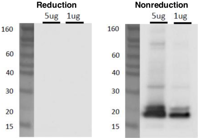 |
| Western blot analysis of CD9 in C32 melanoma cell exosomes using anti-CD9 (12A12) antibody. Sample: C32 cell Exosome (5ug and 1ug) Primary antibody: anti-CD9 (12A12) (1ug/mL) Secondary antibody: anti-mouse IgG-HRP |
|
| Figure 3 | 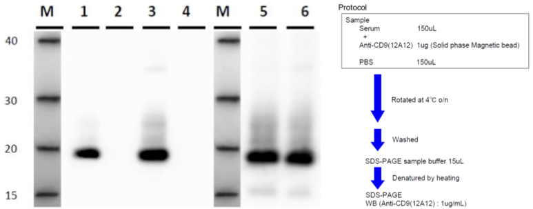 |
| IP-WB of human serum exosomes. Sample: 1. Anti-CD9 (12A12) - Serum A 2. Control antibody - Serum A 3. Anti-CD9 (12A12) - Serum B 4. Control antibody - Serum B 5. Ultracentrifugation - Serum A 6. Ultracentrifugation - Serum B |
|
| Figure 4 | 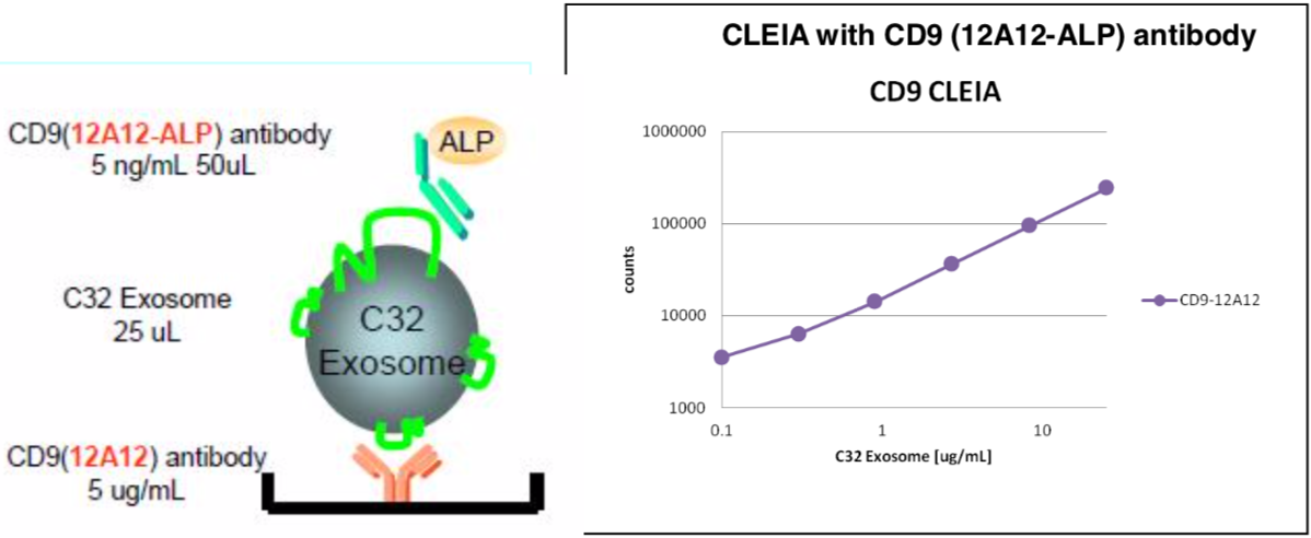 |
| ELISA of C32 melanoma cell exosomes with anti-CD9 (12A12-ALP) antibody (5 ng/mL). | |
| Figure 5 |  |
| Immunoelectron microscopy of exosomes. Extracellular vesicles from human breast cancer cell line (MDA-MB-231-luc-D3H2LN)) were stained with anti-human CD9 antibody (clone 12A12). |
|
| Figure 6 |  |
| ELISA performance of clone 12A12 (anti-CD9) relative to competitor clone A. Clone A failed to generate a signal while clone 12A12 detected a strong signal. |
|
| Product name | Anti CD63 Antigen (LAMP-3/Tspan-30) mAb (Clone 8A12) |
| Cat No | CAC-SHI-EXO-M02-50 |
| Product name | Anti CD63 Antigen (LAMP-3/Tspan-30) mAb (Clone 8A12, Biotin Labeled) |
| Cat No | CAC-SHI-EXO-M02-B |
| Product name | Anti CD63 Antigen (LAMP-3/Tspan-30) (Clone 8A12, TF2 Labeled) |
| Cat No | CAC-SHI-EXO-MO2-TF2 |
| Product name | Anti CD63 Antigen (LAMP-3/Tspan-30) (Clone 8A12, TF5 Labeled) |
| Cat No | CAC-SHI-EXO-M02-TF5 |
| Description | CD63 (also known as LAMP-3, Melanoma-associated antigen ME491, TSPAN30, MLA1 and OMA81H) is a cell surface glycoprotein which belongs to the tetraspanin superfamily. CD63 is known to complex with integrins. CD63 is expressed on activated platelets, monocytes and macrophages, and is weakly expressed on granulocytes, T cells and B cells. Importantly, it is found on the surface of exosomes. Exosomes are cell-derived vesicles bounded by a lipid bilayer membrane and exhibiting a diameter of 50 to 150 nm. They are secreted from almost all cells and are observed in body fluids such as saliva, blood, urine, amniotic fluid, malignant ascites. Recent studies indicate that exosomes contain various proteins and RNAs, suggesting a role in information transfer between cells. This monoclonal antibody can be used to immunoprecipitate exosomes from serum and culture supernatants. References: 1) Yoshioka Y et al., Nat Commun. 2014 Apr 7;5:3591. doi: 10.1038/ncomms4591. 2) N Nishida-Aoki et al., Mol Ther. 2017 Jan 4;25(1):181-191. doi: 10.1016/j.ymthe.2016.10.009. 3) Saito S et al., Sci Rep. 2018 Mar 5;8(1):3997. doi: 10.1038/s41598-018-22450-2. |
| Host | Mouse |
| Species specificity | HU |
| Figure 1 |  |
| Generic exosome shown with molecules commonly found associated with exosomes. Not all molecules will be found with any given exosome and other molecules may also be found. | |
| Figure 2 | 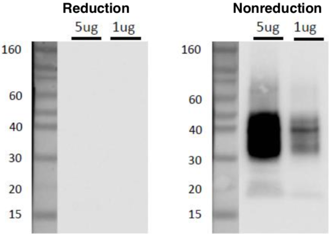 |
| Western blot analysis of CD63 in C32 cell exosomes with anti-CD63 (8A12) antibody. Sample: C32 cell exosomes (5ug and 1ug) Primary antibody: anti-CD63 (8A12) (1ug/mL) Secondary antibody: anti-mouse IgG-HRP |
|
| Figure 3 | 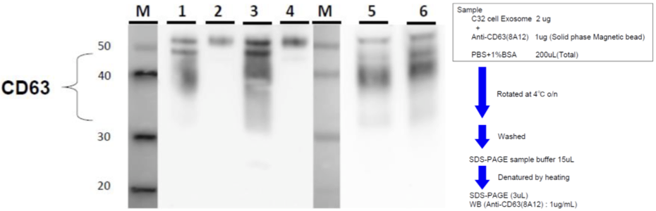 |
| IP-WB of human serum exosomes. Sample 1. Anti-CD63 (8A12) - Serum A 2. Control antibody - Serum A 3. Anti-CD63 (8A12) - Serum B 4. Control antibody - Serum B 5. Ultracentrifugation - Serum A 6. Ultracentrifugation - Serum B |
|
| Figure 4 | 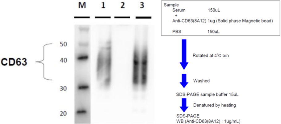 |
| IP-WB of C32 melanoma cell exosomes. Sample 1. Anti-CD63 (8A12) 2. Control antibody 3. C32 cell exosomes (0.4 ug) |
|
| Figure 5 | 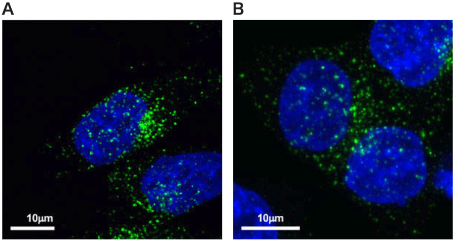 |
| Immunofluorescence localization of CD63 in prostate cancer cell PC-3M and colon cancer cell line HCT116. A: Prostate cancer cell line PC-3MB B: Colon cancer cell line HCT116 Secondary antibody: Alexa Fluor® 488-labeled anti-mouse Data provided: Professor Kosuke Kosaka, Department of Molecular Cell Therapy, National Cancer Center Research Institute, Tokyo Medical University Business-Academia Collaboration Course Extracellular vesicle drug discovery research course. |
|
| Figure 6 |  |
| Immunoelectron microscopy of exosomes. Human breast cancer cell-derived exosomes were stained with anti-human CD63 antibody (clone 8A12). Data provided by: Professor Nao Nishida, National Cancer Center Research Institute Molecular cell therapy research field. |
|
| Figure 7 |  |
| ELISA performance of clone 8A12 (anti-CD63) relative to competitor clone B. Clone 8A12 could detect a stronger signal than clone B. |
|
| Product name | Anti CD81 Antigen (TAPA-1/Tspan-28) mAb (Clone 12C4) |
| Cat No | CAC-SHI-EXO-M03-50 |
| Product name | Anti CD81 Antigen (TAPA-1/Tspan-28) mAb (Clone 12C4, Biotin Labeled) |
| Cat No | CAC-SHI-EXO-M03-B |
| Product name | Anti CD81 Antigen (TAPA-1/Tspan-28) mAb (Clone 12C4, TF2 Labeled) |
| Cat No | CAC-SHI-EXO-MO3-TF2 |
| Product name | Anti CD81 Antigen (TAPA-1/Tspan-28) mAb (Clone 12C4, TF5 Labeled) |
| Cat No | CAC-SHI-EXO-M03-TF5 |
| Description | CD81 (TAPA-1) is a cell surface protein which belongs to the tetraspanin superfamily. CD81 is identified as a component of the B lymphocyte receptor (BCR) and as a receptor for the Hepatitis C Virus. Importantly, it is found on the surface of exosomes. Exosomes are cell-derived vesicles bounded by a lipid bilayer membrane and exhibiting a diameter of 50 to 150 nm. They are secreted from almost all cells and are observed in body fluids such as saliva, blood, urine, amniotic fluid, malignant ascites. Recent studies indicate that exosomes contain various proteins and RNAs, suggesting a role in information transfer between cells. This monoclonal antibody can be used to immunoprecipitate exosomes from serum and culture supernatants. References: 1) Takahashi A et al., Nat Commun. 2017 May 16;8:15287. doi: 10.1038/ncomms15287. 2) M Somiya et al., J Extracell Vesicles. 2018 Feb 21;7(1):1440132. doi:10.1080/20013078.2018.1440132. eCollection 2018. |
| Host | Mouse |
| Species specificity | HU BOV |
| Figure 1 | 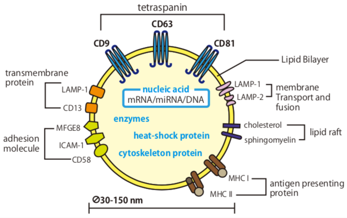 |
| Generic exosome shown with molecules commonly found associated with exosomes. Not all molecules will be found with any given exosome and other molecules may also be found. | |
| Figure 2 | 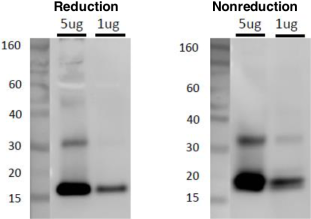 |
| Western blot analysis of CD81 in C32 melanoma cell exosomes with anti-CD81 (clone 12C4) antibody. Sample 1. C32 cell Exosome (5ug, 1ug) 2. Primary antibody: anti-CD81 (clone 12C4) (1ug/mL) 3. Secondary antibody: anti-mouse IgG-HRP |
|
| Figure 3 | 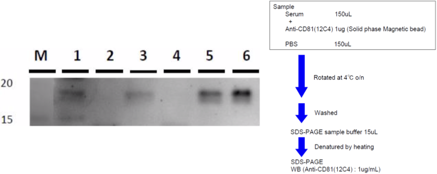 |
| IP-WB of human serum exosomes. Sample 1. Anti-CD81 (12C4) - Serum A 2. Control antibody - Serum A 3. Anti-CD81 (12C4) - Serum B 4. Control antibody - Serum B 5. Ultracentrifugation - Serum A 6. Ultracentrifugation - Serum B |
|
| Figure 4 | 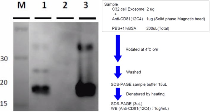 |
| IP-WB of C32 Melanoma cell line exosomes. Sample 1. Anti-CD81 (12C4) 2. Control antibody 3. C32 cell exosomes (0.4 ug) |
|
| Product name | Anti Bovine Milk Exosome pAb (Rabbit, Ammonium Sulfate Purified) |
| Cat No | CAC-EXO-AB-01 |
| Description | Exosomes are membrane vesicles with a diameter of about 50 nm to 150 nm. They are secreted by most cells and are observed in body fluids such as saliva, blood, urine, amniotic fluid and malignant ascites. By encapsulating miRNA, mRNA, protein, micro peptides exosomes may be responsible for transmitting information to target cells or tissues. It is known that cancer metastasis can be suppressed by neutralizing exosome signaling with antibodies specific for exosome surface antigens such as CD9 and CD63. Exosomes are also present in the milk of a wide range of animals such as humans, cows and rats. Furthermore, recent studies have demonstrated the utility of exosomes derived from raw milk as a means of orally administering anticancer agents. References: 1) Hoshino A, et al. (2015) Tumour exosome integrins determine organoorganic metabolism. Nature. 527(7578):329-35. 2) Nao Nishida-Aoki, et al. (2017) Disruption of Circulating Extracellular Vesicles as a Novel Therapeutic Strategy against Cancer Metastasis. Mol Ther. 25(1):181-191. 3) Hirohisa Izumi, et al. (2015) Bovine milk exosomes contain microRNA and mRNA and is taken up by human macrophages. J Dairy Sci. 98(5):2920-33. 4) Ashish K. Agrawal et al. (2017) Milk-derived exosomes for oral delivery of paclitaxel. Nanomedicine. 13(5):1627-1636 |
| Host | RAB |
| Species specificity | BOV |
| Figure 1 | 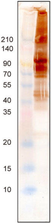 |
| Immunoblot analysis of raw milk with anti-milk exosome antibody (CAC-EXO-AB-01). | |