 CAC Antibody Collection
The antibodies on this page are part of Cosmo Bio's exclusive CAC Collection. For many many thousands of other antibodies from many different makers, use our Search the Store function and our Explore Products drop down menu.
Cytoskeleton
An intriguing feature of eukaryotic cells is the ability of extracts that contain cytosol, devoid of organelles, to roughly maintain the shape of the cell and even to move or contract, depending on how the extracts are pre- pared. This maintenance of structure by the cytosol arises from a complex network of protein filaments that traverse the cell cytoplasm, called the cytoskeleton. The cytoskeleton is not simply a passive feature of the cell that provides structural integrity; it is a dynamic structure that is responsible for whole-cell movement, changes in cell shape, and contraction of muscle cells—it pro- vides the machinery to move organelles from one place to another in the cytoplasm. In addition, recent studies have provided evidence that the cytoskeleton is the master organizer of the cell’s cytoplasm, furnishing binding sites for the specific localization of ribonucleic acids (RNA) and proteins that were once thought to diffuse freely through the cytoplasm.Amazingly, the many activities of the cytoskeleton depend on just three principal types of protein assemblies: actin filaments, microtubules, and intermediate filaments (IFs). Each type of filament or microtubule is formed by a specific association of protein monomers. The dynamic aspects of the cytoskeletal structures arise from accessory proteins that control the length of the assemblies, their position within the cell, and the specific-binding sites along the filaments and microtubules for association with protein complexes, organelles, and the cell membrane. Thus, although the protein filaments and microtubules define the cytoskeleton, the participation of accessory or regulatory proteins conveys its diverse activities. [from: Goodman, Steven R. “Chapter 3 - Cytoskeleton.” Medical Cell Biology (Third Edition). Ed. Steven R. Goodman. Texas: Academic Press, 2008. 59-100. Science Direct. Web. 28 May. 2019.]
| Product name | Anti Keratin, Type I Cytoskeletal 18 (KRT18/Cytokeratin 18) mAb (Clone D2C7) |
| Cat No | CAC-CE-044A |
| Description | The intermediate filaments consist of a large number of nuclear and cytoplasmic proteins that are expressed in a tissue- and differentiation-dependent manner. The components of the intermediate filaments are highly conserved during evolution and show a high degree of conservation among species. Cytokeratins are major structural proteins found in epithelial cells, which form the cytoplasmic network of intermediate filaments. Cytokeratins consist of at least 20 unique gene products that fall into two categories: the relatively acidic type I group (CK9–CK20) and the neutral–basic type II group (CK1–CK8). These cytokeratins are combined in a 1:1 ratio into noncovalent heteropolymers, which are further assembled into keratin filaments. The expression of cytokeratin proteins depends primarily on the epithelial cell type and its degree of differentiation; therefore, assessments of cytokeratin expression status are useful for distinguishing carcinomas from other types of cancer.The cytokeratin 18 gene (CK18) is located on chromo-some 12q13 and is 3,791 base pairs long. It codes for a type I intermediate filament protein that is found primarily in many types of single-layered or "simple" epithelial tissues and is localized in the cytoplasm and perinuclear region.CK18 and its coexpressed complementary type II keratin partner, CK8, are persistently expressed in a variety of adult epithelial organs, such as the liver, lung, kidney, pancreas, gastrointestinal tract, and mammary gland, and are also expressed by cancers that arise from these tissues. In the absence of CK8, the CK18 protein is degraded and keratin intermediate filaments are not formed. A cDNA clone forCK8 was isolated and expressed in mouse fibroblasts that had previously been transfected with the CK18 gene, which resulted in the formation of stable keratin filaments.Thus, the presence of a type I (CK18) and a type II (CK8)keratin appears to be both necessary and sufficient for the formation of keratin filaments. [from: Yu-Rong Weng Y-R., Cui Y. and Fang J-Y. (2012) Biological Functions of Cytokeratin 18 in Cancer. Mol Cancer Res 10(4):485-493; DOI: 10.1158/1541-7786.MCR-11-0222] |
| Host | MS |
| Species specificity | HU MS RT MKY |
| Figure 1 | 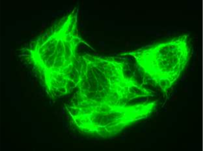 |
| Immunocytochemistry / Immunofluorescence analysis of Cytokeratin-18 (CK18) antibody (D2C7) in HeLa cells. | |
| Figure 2 | 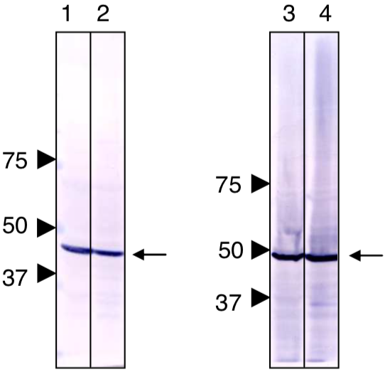 |
| Immunoblot analysis of Cytokeratin-18 (CK18) antibody (D2C7). 1.HeLa cell extract. 2.COS1 cell extract. 3.rat liver extract. 4. mouse liver extract. |
|
| Product name | Anti Tubulin Gamma-1 Chain (GCP-1) mAb (Clone E39) |
| Cat No | CAC-NM-MA-003 |
| Description | Gamma-tubulins constitute a ubiquitous and highly conserved subfamily of the tubulin family. The protein is found at microtubule-organizing centers, such as spindle poles or centrosomes. It remains associated with centrosomes when microtubules are depolymerized, suggesting that it is an integral component that may play a role in minus-end nucleation of microtubule assembly. Thus, gamma-tubulins can be used as a centrosome maker. |
| Host | MS |
| Species specificity | HU MS RT |
| Figure 1 | 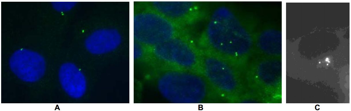 |
| Immunofluorescence analysis of monoclonal antibody (E39) recognition of gamma-tubulin at centrosomes. Gamma-tubulin in normal or cancer cells was stained by immunofluorescence described below. The presence of supernumerary centrosomes is a common event in human tumors. Yellow and blue indicate gamma-tubulin and nuclear DNA, respectively. A: normal human fibroblasts. B: colon cancer cell line (CaCo-2). C: fibrosarcoma cell line (HT-1080). |
|