
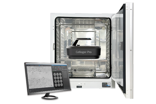
Celloger® Pro Automated Live Cell Imaging System
Curiosis
- Catalog No.:
- QRI-CRCLG-P01


Please contact us to request a pricing quote
Related Products

Product Description
Celloger® Pro is an advanced live cell imaging system offering exceptional image quality and convenience. It enables real-time cell monitoring inside the incubator, allowing seamless observation and tracking of cellular dynamics without disturbing the natural growth environment. With dual fluorescence and bright-field microscopy, multiple markers can be visualized simultaneously, while multi-point time-lapse imaging captures dynamic cellular events at various locations.
Key Features
• Real-time cell monitoring inside an incubator
• Dual fluorescence microscopy for enhanced imaging
• Multi-point time-lapse imaging capability
• User-interchangeable objective lens option
• Intuitive interface and user-friendly tools
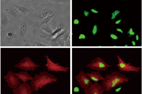
Multicolor fluorescence and brightfield imaging
With its dual color fluorescence and bright-field imaging capabilities, Celloger® Pro enables the capture of high-quality and high-resolution images.
With enhanced scanning methods and innovative merging techniques, the system reduces scanning time, enabling researchers to analyze cellular dynamics with exceptional clarity and efficiency.
Real-time monitoring inside the incubator
Celloger® Pro is designed to facilitate real-time monitoring of cells inside an incubator. By simply placing the device within the incubator and connecting it to an external PC, researchers are able to remotely observe cells in real-time.
With the time-lapse function, cell images are captured according to the schedule set by the researcher; the images can then be easily converted into time-lapse videos.
User-interchangeable objective lens
Celloger® Pro offers user-interchangeable objective lenses, providing flexibility to researchers based on their specific study requirements. With options such as 2X, 4X, and 10X objectives, users can switch between these lenses by hand.
Purchase interchangeable objective lenses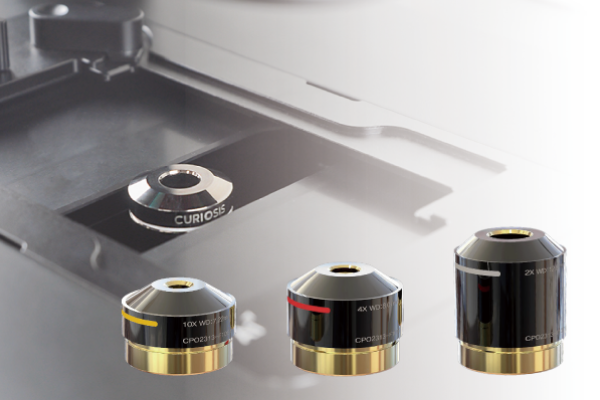
Capturing images from multiple positions
Celloger® Pro enables imaging of samples in multiple positions by automatically moving the integrated camera while keeping the vessel and sample fixed on the stage. This ensures a stable environment for the cells, resulting in enhanced image quality and precise research outcomes.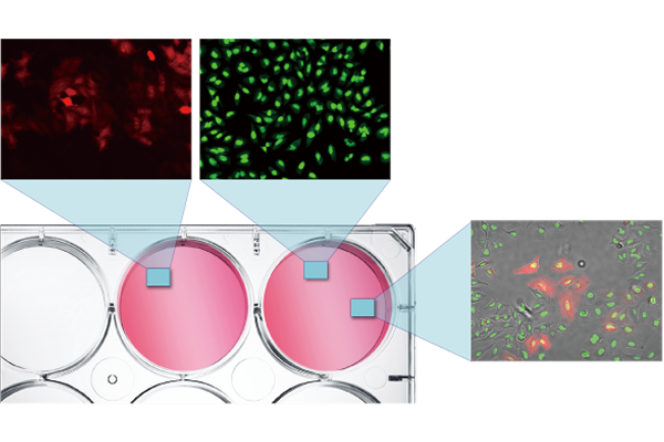
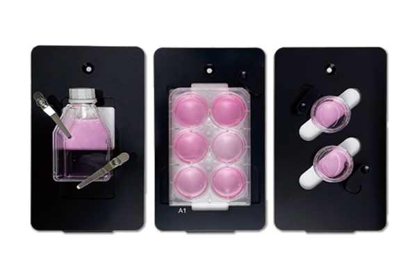
Compatible with different vessel types
The system is compatible with different cell culture vessels such as well plates (up to 96 wells), flasks, dishes, and slides, and can switch between them by simply replacing the vessel holders for specific needs.
Application Videos
Celloger® Pro Application I Actin dynamics (Depolymerization)
HeLa cells expressing tdTomato-actin were treated with 1.25 μM and 10 μM cytochalasin B, with 1 hour interval images taken over 48 hours.
Celloger® Pro Application I Adipogenesis
Observe adipocyte differentiation in 3T3-L1 cells stained with LipiDye II over 48 hours, capturing 1-hour interval images.
Celloger® Pro Application I NK cell killing assay
The U-2OS cells(target) were stained with CellTracker Green CMFDA and then co-cultured with NK-92 cells to observe the process of NK cells killing the target.
Celloger® Pro Application I Spheroid cytotoxicity
Monitor the drug effect on HEK293-GFP spheroids treated with Staurosporine by capturing images every 30 minutes for 24 hours.
Celloger® Pro Application I Transfection efficiency
To monitor gene transfection efficiency in real-time, AGS cells were stained with CMFDA dye and then transfected with the tdTomato-Lifeact gene.
Concentration: >1x10¹⁰ particles/mL
Specifications
|
Imaging modes
|
Brightfield, Dual fluorescence (Green & Red) |
| Fluorescence | Green : EX (470/40), EM (540/50) Red : EX (562/40), EM (641/75) |
| Camera | High sensitivity 5.0 MP CMOS |
| Field of view | 2X (2.02 x 1.49 mm) / 4X (1.41 x 1.05 mm) / 10X (0.70 x 0.52 mm) |
| Imaging methods | Single/multicolor, stitching, stacking, time-lapse, real-time recording |
| Dimensions (H x W x L) | 250 x 338 x 412 mm |
| Culture vessels | Well plate up to 96-well, flask, dish, slide |
| Operating environment | 10~40℃, 20~95% humidity |
| O/S required | Windows 10 and above |
| Objective Lens | 2X, 4X, 10X (User-interchangeable) |
| Stage | Fully motorized XYZ (Fixed stage, camera moving type) |
| Imaging positions | Multiple |
| Focus | Autofocus, Manual focus |
| Included software | Scan App, Analysis App |
| Weight | 9.6 kg |
| File export format | TIFF, AVI (JPEG, PNG) |
| Power requirement | 100-240V, ~50/60Hz |
| Incubator specification | Above 200L (recommended)
|
| Documents & Links for Celloger® Pro Automated Live Cell Imaging System | |
| Datasheet | Celloger® Pro Automated Live Cell Imaging System Datasheet |
| Documents & Links for Celloger® Pro Automated Live Cell Imaging System | |
| Datasheet | Celloger® Pro Automated Live Cell Imaging System Datasheet |