Anti VDR mAb (Clone 3C7)

CUSABIO

SKU:
CSB-RA945260A0HU-50
  • Western Blot<br />
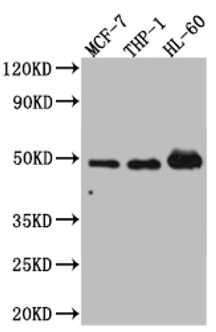
Positive WB detected in: MCF-7 whole cell lysate, THP-1 whole cell lysate, HL-60 whole cell lysate<br />
All lanes: Vitamin D antibody at 1:1000<br />
Secondary<br />
Goat polyclonal to rabbit IgG at 1/50000 dilution<br />
Predicted band size: 49, 54 kDa<br />
Observed band size: 49 kDa<br />
  • IHC image of CSB-RA945260A0HU diluted at 1:100 and staining in paraffin-embedded human colon cancer performed on a Leica BondTM system. After dewaxing and hydration, antigen retrieval was mediated by high pressure in a citrate buffer (pH 6.0). Section was blocked with 10% normal goat serum 30min at RT. Then primary antibody (1% BSA) was incubated at 4℃ overnight. The primary is detected by a Goat anti-rabbit IgG polymer labeled by HRP and visualized using 0.05% DAB.
Shipping:
Calculated at Checkout
$221.00
Adding to cart… The item has been added
Nuclear hormone receptor. Transcription factor that mediates the action of vitamin D3 by controlling the expression of hormone sensitive genes. Recruited to promoters via its interaction with BAZ1B/WSTF which mediates the interaction with acetylated histones, an essential step for VDR-promoter association. Plays a central role in calcium homeostasis.

Product Specifications
Application WB, IHC, ELISA
Reactivity Human
Clonality Monoclonal (Clone No.: 3C7)


Documents & Links for Anti VDR mAb (Clone 3C7)
Datasheet Anti VDR mAb (Clone 3C7) Datasheet

Documents & Links for Anti VDR mAb (Clone 3C7)
Datasheet Anti VDR mAb (Clone 3C7) Datasheet