Anti MAPT mAb (Clone 4A1)

CUSABIO

SKU:
CSB-RA246354A0HU-50
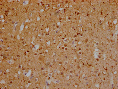
  • IHC image of CSB-RA246354A0HU diluted at 1:100 and staining in paraffin-embedded human glioma cancer performed on a Leica BondTM system. After dewaxing and hydration, antigen retrieval was mediated by high pressure in a citrate buffer (pH 6.0). Section was blocked with 10% normal goat serum 30min at RT. Then primary antibody (1% BSA) was incubated at 4℃ overnight. The primary is detected by a Goat anti-rabbit IgG polymer labeled by HRP and visualized using 0.05% DAB.
  • IHC image of CSB-RA246354A0HU diluted at 1:100 and staining in paraffin-embedded human brain tissue performed on a Leica BondTM system. After dewaxing and hydration, antigen retrieval was mediated by high pressure in a citrate buffer (pH 6.0). Section was blocked with 10% normal goat serum 30min at RT. Then primary antibody (1% BSA) was incubated at 4℃ overnight. The primary is detected by a Goat anti-rabbit IgG polymer labeled by HRP and visualized using 0.05% DAB.
Shipping:
Calculated at Checkout
$221.00
Adding to cart… The item has been added
Promotes microtubule assembly and stability, and might be involved in the establishment and maintenance of neuronal polarity. The C-terminus binds axonal microtubules while the N-terminus binds neural plasma membrane components, suggesting that tau functions as a linker protein between both. Axonal polarity is predetermined by TAU/MAPT localization (in the neuronal cell) in the domain of the cell body defined by the centrosome. The short isoforms allow plasticity of the cytoskeleton whereas the longer isoforms may preferentially play a role in its stabilization.

Product Specifications
Application IHC, ELISA
Reactivity Human
Clonality Monoclonal (Clone No.: 4A1)


Documents & Links for Anti MAPT mAb (Clone 4A1)
Datasheet Anti MAPT mAb (Clone 4A1) Datasheet

Documents & Links for Anti MAPT mAb (Clone 4A1)
Datasheet Anti MAPT mAb (Clone 4A1) Datasheet