Anti HSPD1 mAb (Clone 3D8)

CUSABIO

SKU:
CSB-RA953395A0HU-50
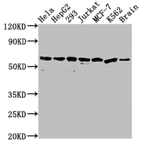
  • Western Blot<br />
Positive WB detected in: Hela whole cell lysate, HepG2 whole cell lysate, 293 whole cell lysate, Jurkat whole cell lysate, MCF-7 whole cell lysate, K562 whole cell lysate, Mouse brain tissue<br />
All lanes: HSPD1 antibody at 1:2000<br />
Secondary<br />
Goat polyclonal to rabbit IgG at 1/50000 dilution<br />
Predicted band size: 62, 18 kDa<br />
Observed band size: 60 kDa<br />
  • IHC image of CSB-RA953395A0HU diluted at 1:100 and staining in paraffin-embedded human liver tissue performed on a Leica BondTM system. After dewaxing and hydration, antigen retrieval was mediated by high pressure in a citrate buffer (pH 6.0). Section was blocked with 10% normal goat serum 30min at RT. Then primary antibody (1% BSA) was incubated at 4℃ overnight. The primary is detected by a Goat anti-rabbit IgG polymer labeled by HRP and visualized using 0.05% DAB.
  • IHC image of CSB-RA953395A0HU diluted at 1:100 and staining in paraffin-embedded human liver cancer performed on a Leica BondTM system. After dewaxing and hydration, antigen retrieval was mediated by high pressure in a citrate buffer (pH 6.0). Section was blocked with 10% normal goat serum 30min at RT. Then primary antibody (1% BSA) was incubated at 4℃ overnight. The primary is detected by a Goat anti-rabbit IgG polymer labeled by HRP and visualized using 0.05% DAB.
  • Immunoprecipitating HSPD1 in HepG2 whole cell lysate<br />
Lane 1: Rabbit control IgG instead of CSB-RA953395A0HU in HepG2 whole cell lysate.
For western blotting,a HRP-conjugated Protein G antibody was used as the secondary antibody (1/2000)<br />
Lane 2: CSB-RA953395A0HU(3µg)+ HepG2 whole cell lysate(500µg)<br />
Lane 3: HepG2 whole cell lysate (10µg)<br />
Shipping:
Calculated at Checkout
$221.00
Adding to cart… The item has been added
Chaperonin implicated in mitochondrial protein import and macromolecular assembly. Together with Hsp10, facilitates the correct folding of imported proteins. May also prevent misfolding and promote the refolding and proper assembly of unfolded polypeptides generated under stress conditions in the mitochondrial matrix (PubMed:1346131, PubMed:11422376). The functional units of these chaperonins consist of heptameric rings of the large subunit Hsp60, which function as a back-to-back double ring. In a cyclic reaction, Hsp60 ring complexes bind one unfolded substrate protein per ring, followed by the binding of ATP and association with 2 heptameric rings of the co-chaperonin Hsp10. This leads to sequestration of the substrate protein in the inner cavity of Hsp60 where, for a certain period of time, it can fold undisturbed by other cell components. Synchronous hydrolysis of ATP in all Hsp60 subunits results in the dissociation of the chaperonin rings and the release of ADP and the folded substrate protein (Probable).

Product Specifications
Application WB, IHC, IP, ELISA
Reactivity Human, Mouse
Clonality Monoclonal (Clone No.: 3D8)


Documents & Links for Anti HSPD1 mAb (Clone 3D8)
Datasheet Anti HSPD1 mAb (Clone 3D8) Datasheet

Documents & Links for Anti HSPD1 mAb (Clone 3D8)
Datasheet Anti HSPD1 mAb (Clone 3D8) Datasheet