Anti FOS mAb (Clone 14C10)

CUSABIO

SKU:
CSB-RA008790A0HU-50
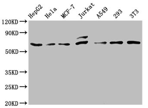
  • Western Blot<br />
Positive WB detected in: HepG2 whole cell lysate, Hela whole cell lysate, MCF-7 whole cell lysate, Jurkat whole cell lysate, A549 whole cell lysate, 293 whole cell lysate, NIH/3T3 whole cell lysate<br />
All lanes: c-FOS antibody at 0.81μg/ml<br />
Secondary<br />
Goat polyclonal to rabbit IgG at 1/50000 dilution<br />
Predicted band size: 41, 29, 37 KDa<br />
Observed band size: 62 KDa<br />
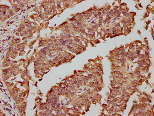
  • IHC image of CSB-RA008790A0HU diluted at 1:81 and staining in paraffin-embedded human adrenal gland tissue performed on a Leica BondTM system. After dewaxing and hydration, antigen retrieval was mediated by high pressure in a citrate buffer (pH 6.0). Section was blocked with 10% normal goat serum 30min at RT. Then primary antibody (1% BSA) was incubated at 4℃ overnight. The primary is detected by a biotinylated secondary antibody and visualized using an HRP conjugated SP system.
  • IHC image of CSB-RA008790A0HU diluted at 1:81 and staining in paraffin-embedded human cervical cancer performed on a Leica BondTM system. After dewaxing and hydration, antigen retrieval was mediated by high pressure in a citrate buffer (pH 6.0). Section was blocked with 10% normal goat serum 30min at RT. Then primary antibody (1% BSA) was incubated at 4℃ overnight. The primary is detected by a biotinylated secondary antibody and visualized using an HRP conjugated SP system.
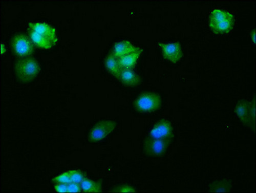
  • Immunofluorescence staining of HepG2 cells with CSB-RA008790A0HU at 1:27, counter-stained with DAPI. The cells were fixed in 4% formaldehyde, permeabilized using 0.2% Triton X-100 and blocked in 10% normal Goat Serum. The cells were then incubated with the antibody overnight at 4℃. The secondary antibody was Alexa Fluor 488-congugated AffiniPure Goat Anti-Rabbit IgG (H+L).
Shipping:
Calculated at Checkout
$221.00
Adding to cart… The item has been added
Nuclear phosphoprotein which forms a tight but non-covalently linked complex with the JUN/AP-1 transcription factor. In the heterodimer, FOS and JUN/AP-1 basic regions each seems to interact with symmetrical DNA half sites. On TGF-beta activation, forms a multimeric SMAD3/SMAD4/JUN/FOS complex at the AP1/SMAD-binding site to regulate TGF-beta-mediated signaling. Has a critical function in regulating the development of cells destined to form and maintain the skeleton. It is thought to have an important role in signal transduction, cell proliferation and differentiation. In growing cells, activates phospholipid synthesis, possibly by activating CDS1 and PI4K2A. This activity requires Tyr-dephosphorylation and association with the endoplasmic reticulum.

Product Specifications
Application WB, IHC, IF, FC, ELISA
Reactivity Human, Mouse
Clonality Monoclonal (Clone No.: 14C10)


Documents & Links for Anti FOS mAb (Clone 14C10)
Datasheet Anti FOS mAb (Clone 14C10) Datasheet

Documents & Links for Anti FOS mAb (Clone 14C10)
Datasheet Anti FOS mAb (Clone 14C10) Datasheet