Color changes depending on the polarity of the environment such as intracellular organelles
POLARIC™ Live Cell Staining Fluorescent Dye
POLARIC™, a fluorescent solvatochromic dye using the Suzuki cross-coupling method, changes its color (fluorescence wavelength) depending on the polarity of the environment (new water/hydrophobicity) such as subcellular organelles with a single excitation light. .
Intracellular organelles can be dyed separately. Please use it for different staining according to cell types, imaging of phenotypic changes in living cells, etc.
*This product is manufactured and sold by Cosmo Bio under a license agreement with Hokkaido University.
Features
- POLARIC™ changes color according to the polarity (hydrophilic/hydrophobic) of the environment such as intracellular organelles
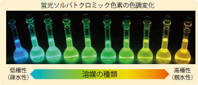
POLARIC™, a fluorescent solvatochromic dye, changes its fluorescence wavelength depending on the polarity of the solvent.
POLARIC™ has a wide range of emission wavelengths from approximately 520 to 700 nm with excitation light around 500 nm. - POLARIC™ has extremely low cytotoxicity and long fluorescence lifetime
Dyeing method
- Add 3 μL of ethanol to a POLARIC™ 1 tube to dissolve, and add to 10 mL of medium to make the staining solution.
Store the staining solution at 4°C for 2 weeks (protected from light). - Incubate the target cells in a culture vessel with low autofluorescence such as a “glass bottom dish”.
- Remove the medium from the culture vessel and replace with an equal volume of staining solution.
*We recommend staining at the early stages of culture. - Allow to stand in a culture environment (37°C incubator, etc.) for staining.
Standard staining time is about 10-30 minutes for floating cells and cells before seeding, and about 15 minutes-2 hours (up to 12 hours) for cells after seeding.
*Staining time varies depending on the type of cell and number of culture days. - Wash three times with medium after staining.
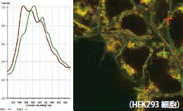
- Fluorescently observe cells after washing. The observation conditions are an excitation wavelength of 500 nm (460 nm to 520 nm) and an emission wavelength of approximately 520 to 700 nm (depending on the localized environment within the cell).
The main cell organelles stained with POLARIC™ are the cell membrane (green), mitochondria (orange), and endoplasmic reticulum (yellow).
Example of use
POLARIC™ changes color depending on the polarity of the intracellular site.
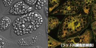
Mitochondria fluoresce orange, cell membranes green, and endoplasmic reticulum yellow.
Phase contrast image
nuclear staining
POLARIC™ staining
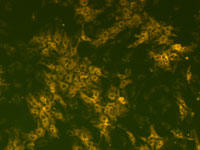
Rat cardiomyocytes stained with POLARIC™ PLT-500c6 are able to differentiate between cardiomyocytes and non-cardiomyocytes. Even stained cardiomyocytes continue to beat. Check out the video here .
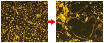
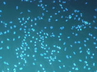
When rat bone marrow monocytes stained with POLARIC™ are cultured in osteoclast differentiation medium, it is possible to observe the progress of differentiation into osteoclasts while remaining stained.
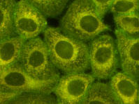
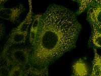
HeLa cells stained with POLARIC™ PLT-500c6
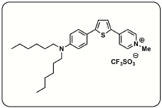
Structure of POLARIC™ PLT-500c6
| Catalog Number | Product Name | Size |
| PMC-AK12 | POLARIC PLT-500c6 | 5 Tubes |