Anti ATP5A1 mAb (Clone 5G11)

CUSABIO

SKU:
CSB-RA159926A0HU-50
  • Western Blot<br />
Positive WB detected in: HepG2 whole cell lysate, MCF-7 whole cell lysate, 293 whole cell lysate, HL60 whole cell lysate, Mouse Heart tissue, Mouse Brain tissue<br />
All lanes: ATP5F1A antibody at 1:2000<br />
Secondary<br />
Goat polyclonal to rabbit IgG at 1/50000 dilution<br />
Predicted band size: 60, 55, 58 kDa<br />
Observed band size: 60 kDa<br />
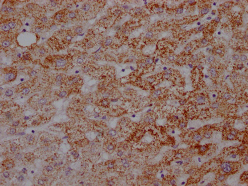
  • IHC image of CSB-RA159926A0HU diluted at 1:100 and staining in paraffin-embedded human liver tissue performed on a Leica BondTM system. After dewaxing and hydration, antigen retrieval was mediated by high pressure in a citrate buffer (pH 6.0). Section was blocked with 10% normal goat serum 30min at RT. Then primary antibody (1% BSA) was incubated at 4℃ overnight. The primary is detected by a Goat anti-rabbit IgG polymer labeled by HRP and visualized using 0.05% DAB.
  • IHC image of CSB-RA159926A0HU diluted at 1:100 and staining in paraffin-embedded human kidney tissue performed on a Leica BondTM system. After dewaxing and hydration, antigen retrieval was mediated by high pressure in a citrate buffer (pH 6.0). Section was blocked with 10% normal goat serum 30min at RT. Then primary antibody (1% BSA) was incubated at 4℃ overnight. The primary is detected by a Goat anti-rabbit IgG polymer labeled by HRP and visualized using 0.05% DAB.
Shipping:
Calculated at Checkout
$221.00
Adding to cart… The item has been added
Mitochondrial membrane ATP synthase (F(1)F(0) ATP synthase or Complex V) produces ATP from ADP in the presence of a proton gradient across the membrane which is generated by electron transport complexes of the respiratory chain. F-type ATPases consist of two structural domains, F(1) - containing the extramembraneous catalytic core, and F(0) - containing the membrane proton channel, linked together by a central stalk and a peripheral stalk. During catalysis, ATP synthesis in the catalytic domain of F(1) is coupled via a rotary mechanism of the central stalk subunits to proton translocation. Subunits alpha and beta form the catalytic core in F(1). Rotation of the central stalk against the surrounding alpha(3)beta(3) subunits leads to hydrolysis of ATP in three separate catalytic sites on the beta subunits. Subunit alpha does not bear the catalytic high-affinity ATP-binding sites (By similarity).

Product Specifications
Application WB, IHC, ELISA
Reactivity Human, Mouse
Clonality Monoclonal (Clone No.: 5G11)


Documents & Links for Anti ATP5A1 mAb (Clone 5G11)
Datasheet Anti ATP5A1 mAb (Clone 5G11) Datasheet

Documents & Links for Anti ATP5A1 mAb (Clone 5G11)
Datasheet Anti ATP5A1 mAb (Clone 5G11) Datasheet