Other related information: How to use biotin-labeled lectin
How to use biotin-labeled lectins Use for staining glycoproteins on membranes
Features
・Easy operation
・Multiple samples and multiple types of lectins can be stained at once
・Purified samples are required
Method
1. Sample preparation: After dissolving the sample in PBS*1 (concentration 1 mg/ml), prepare a 2-fold dilution series.
2. Blotting onto membrane: Set the membrane *3 on the dot blotting device *2. 30 to 40 μl of sample is placed on the well and allowed to stand at room temperature for 15 minutes.
3. Washing: Vacuum filter samples and add 50 μl of PBS directly to each well. After repeating this process twice, remove the membrane from the dot blotting device while filtering by suction. (Hereafter, common procedure for dot blot and lectin blot)
4. Blocking of membrane: Immerse the membrane in blocking buffer*5 and gently stir at room temperature for 10 minutes. Change the blocking buffer and repeat two more times.
5. Biotin-labeled lectin reaction: Immerse the blocked membrane in biotin-labeled lectin solution (2-5 μg /ml, diluted with blocking buffer). Stir gently at room temperature for 1 hour.
6. Washing: Discard the biotin-labeled lectin solution, soak in blocking buffer, and gently agitate for 10 minutes at room temperature. Change the blocking buffer and repeat two more times.
7. HRP-labeled avidin reaction: Immerse the washed membrane in HRP-labeled avidin solution (about 1000-fold dilution with blocking buffer). Stir gently at room temperature for 30 minutes.
8. Washing: Discard the HRP-labeled avidin solution, soak in blocking buffer and gently agitate for 10 minutes at room temperature. Change the blocking buffer and repeat two more times.
9. Coloring: Immerse in coloring solution*6 (at room temperature for 1-10 minutes). Discard the coloring solution and wash with pure water to stop the reaction.
10. Drying: Membranes should be air dried and stored.
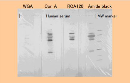
Sample: 5 μl of 50- fold dilution of healthy human serum Amide black
*1 PBS: Phosphate-buffered saline (pH7.2)
*2 Dot blotting device: Biodot (Bio-Rad), dot blot replicator (ATTO), etc.
*3 Membrane: Nitrocellulose (NC) membrane: pure Soak in water for 1 minute before use. Polyvinylidene difluoride (PVDF) membrane: After soaking in methanol for 20 seconds, immerse in pure water for 15 minutes with gentle stirring before use.
*4 Blotting buffer: 25mM Tris, 192mM Glycine, 20% methanol
*5 Blocking buffer: 10mM Tris-HCl pH7.4 + 0.15M NaCl (0.5M NaCl for WGA) + 0.05% Tween20
*6 Coloring solution: POD immunostain Kit (Wako Pure Chemical Industries), 0.03% DAB (diaminobenzidine)/PBS + 0.003% H2O2, etc. 2 / 3

Lectin Blot Method
Features
- Small amount of sample is enough
- Unpurified samples can be used
Method
1. Preparation of sample: Prepare the sample with PBS*1 etc. to an appropriate concentration. Use higher concentrations for unpurified samples. (About 50 times for serum)
2. Electrophoresis
3. Blotting onto membrane: Equilibrate membrane*3 with blotting buffer*4 and attach gel to electrotransfer.
(Hereinafter, the dot blot method)
4. Membrane blocking (same procedure as above)
Biotin-Con A Biotin-WGA Biotin-PHA-E4 Each protein amount is 90μg for No.1.
No.11 is 1.5ng in the following 3-fold dilution series. No.12 is blank.
Figure 2 Results of lectin staining by dot blot method
Usage with Tissue Staining
Preparation of tissue samples
1. Cryosections: PLP (periodic acid-lysine-paraformaldehyde) fixation PFA (paraformaldehyde) fixation
2. Paraffin section: After deparaffinization, 0.3% H2O methanol treatment (removal of endogenous peroxidase)
Dyeing operation
1. Immerse the specimen in PBS*1.
2. Immerse in biotin-labeled lectin solution (5-10 μg /ml, diluted with PBS).
3. Wash with PBS.
4. Immerse in HRP-labeled avidin solution.
5. Wash with PBS.
6. Immerse in coloring solution*2.
7. Wash with pure water.
8. After nuclear staining, dehydrate and mount.
*1 PBS: Phosphate-buffered saline (pH7.2) *2 Chromogenic solution: 0.03% DAB (diaminobenzidine)/50mM Tris-HCl pH7.4 + 0.005% H2O2 For frozen sections, add 10mM NaN3.
Application to Cell Sorters
SBA Transferrin Fetuin Ovalbumin Thyroglobulin BSA 3 / 3 Application to cell sorter 7) 8) 9) Fluorescein activated cell sorter (FACS) is a system for rapidly isolating specific cells. It is a method that combines a specific antibody and FACS, and is widely applied to the tracking of cell differentiation/abnormalities and the separation of genetically modified materials. Replacing the specific antibody with a lectin enables glycan-specific separation of cells.
Method
1. A single suspension of cells (2×10 7 cells/ml) is suspended in buffer (0.1% BSA, 0.02% NaN3/PBS).
2. Add biotin-labeled lectin (5-40 μg /ml).
3. Cells are washed 3 times with buffer.
4. FITC-labeled avidin (20 μg /ml) is added.
*When double staining, use Phycoerythrin-labeled avidin (5 μg/ml) and then incubate with FITC-labeled Anti-Thy-1. (for T cells)
5. Cells are washed 3 times with buffer.
Use in ELISA
Quantification of specific proteins in microplates. It is common practice to quantify and measure binding constants using antibodies.
By converting the antibody to a lectin, it is possible to obtain not only quantification but also information on sugar chains.
References
1) Chadli, A., Caron, M., Joubert, R., Bladier, D., Kocourek, J., Anal. Biochem., 204(1), 198 (1992)
2) Seo, Y., Takahama, K., Res. Pract. Forens. Med. (法医学の実際と研究), 37, 155 (1994)
3) Uehara, F., Sameshima, M., Unoki, K., Okubo, A., Yanagita, T., Sugata, M., Iwakiri, N., Ohba, N., Jpn.J.Ophthalmol., 38(4), 364 (1994)
4) Suzuki, K., Ogawa, K., Taniguchi, K., 味と匂のシンポジウム論文集, 27, 49 (1994)
5) Ueda, T., et al., Okajimas Folia. Anal. Jpn., 73, 325 (1997)
6) Takada, K., et al., 細胞工学別冊「グライコバイオロジー実験プロトコール」, P.236-240 (1996)
7) Imai, Y., et al., Mol. Immunol., 25, 419 (1988)
8) Yamashita, Y., et al., Mol. Immunol., 26, 905 (1989)
9) Takano, M., et al., Jpn. J. Cancer Res., 80, 1228 (1989)
10) Duk, M., Lisowska, E., Wu, J. H., Wu, A. M., Anal. Biochem., 221(2), 266 (1994)
11) Mengeling, B. J., Smith, P. L., Baenziger, J. U., Anal. Biochem., 199(2), 286 (1991)
12) Keusch, J., et al., Clinica. Chimica. Acta, 252, 147 (1996)

