- Product Table Navigator
Nichirei Products Dashboard Page
Product Lineup
Simple stain Kits / Mousestain Kits
High stain Kits
Enzyme substrates
ALK Detection System - Nichirei Products Catalog and Brochures
Brochures
Nichirei N-Histofine Catalog
Download Nichirei Product Catalog (pdf) - References and Technical Information
![]()

Peroxidase and Alkaline Phosphatase Substrate Kits
| Catalog Number | Product Name | Size |
| NIC-425312F | N-Histofine DAB-2V | 500 Tests |
| NIC-425312F | N-Histofine DAB-2V | 1500 Tests |
| NIC-415192F | N-Histofine DAB-3S Kit | 500 Tests |
| NIC-415192F | N-Histofine DAB-3S Kit | 1500 Tests |
| NIC-415161F | N-Histofine New Fuchsin Substrate Kit DISCONTINUED | 2000 Tests |
| NIC-415182F | N-Histofine Simple Stain AEC Solution | 500 Tests |
| NIC-415182F | N-Histofine Simple Stain AEC Solution | 1500 Tests |
Enzyme Substrate Systems
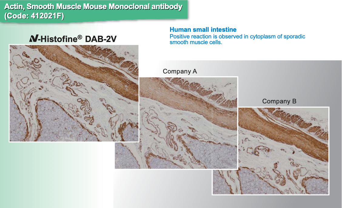
N-Histofine® DAB-2V Kit
N-Histofine® DAB-2V Kit is the updated version of the legacy DAB-3S Kit used to prepare a chromogen-substrate for peroxidase (PO)-based immunohistochemical staining. DAB (3,3’-Diaminobenzidine tetrahydrochloride) produces brown precipitates at target antigen sites reacting with peroxidase.
Advantages
1) Simple to prepare substrate solution
2) Higher sensitivity than other DAB kits
3) Prepared substrate solution stable for two weeks
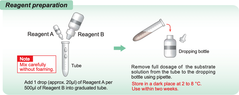
Reagent Preparation
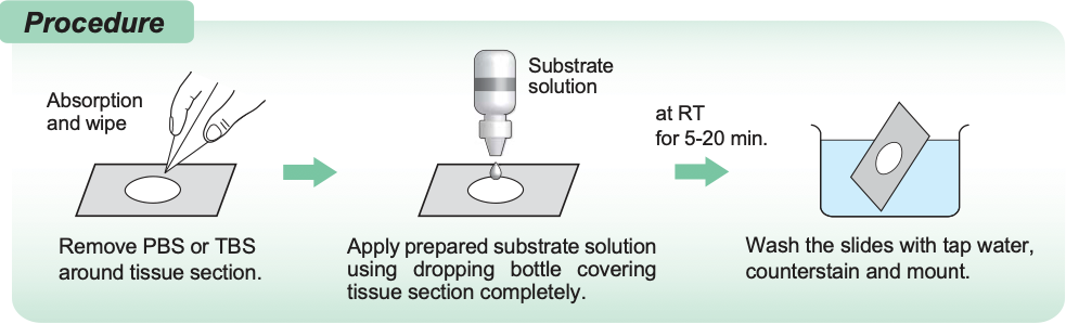
Procedure
Representative Staining Results

INTENDED FOR RESEARCH USE ONLY
N-Histofine® DAB-3S Kit
N-Histofine® DAB-3S Kit is the legacy version of the newly-updated DAB-2V Kit used to prepare a chromogen-substrate for peroxidase (PO)-based immunohistochemical staining. DAB (3,3’-Diaminobenzidine tetrahydrochloride) produces brown precipitates at target antigen sites reacting with peroxidase.
INTENDED FOR RESEARCH USE ONLY
N-Histofine® Simple Stain AEC Solution
N-Histofine® Simple Stain AEC Solution is a pre-made chromogen-substrate solution for peroxidase (PO)-based immunohistochemical staining. AEC (aminoethyl carbazole) produces red precipitates at target antigen sites reacting with peroxidase.
Advantages
1) Ready to use solution requires no advance preparation
2) Solution stable for 18 months after manufacture if stored properly
INTENDED FOR RESEARCH USE ONLY
N-Histofine® New Fuchsin substrate kit DISCONTINUED
N-Histofine® New Fuchsin substrate kit is used to prepare a chromogen-substrate for alkaline phosphatase (AP)-based immunohistochemical staining. New Fuchsin produces red precipitates at target antigen sites reacting with AP.
INTENDED FOR RESEARCH USE ONLY
ALK Detection System
N-Histofine® ALK Detection Kit
N-Histofine® ALK Detection Kit is an immunohistochemical kit designed for convenient, highly-sensitive, and reproducible detection of Anaplastic lymphoma kinase (ALK) proteins expressed in tumor cells present in formalin-fixed paraffin-embedded (FFPE) human tissue sections. Convenient ALK Control Slides are available separately.
Background
The ALK gene located at 2p23 encodes a receptor-type tyrosine kinase belonging to the insulin receptor family. In 1994 the t(2;5)(p23;q35) translocation in anaplastic large-cell lymphoma (ALCL) was shown to result in a fusion between the ALK and NPM (Nucleophosmin) genes. A kinase domain in the ALK protein intracellular region is associated with cell growth promotion and apoptosis inhibition. The proteins produced from ALK fusion genes are chronically activated by dimerization, leading to the development of cancer. Subsequent work has shown that the ALK gene forms fusions with a large number of genes. These include for ALCL: ATIC, CLTC, MSN, TPM3, TPM4, TFG, MYH9 and ALO17; and for inflammatory myofibroblastic tumors (IMT): ATIC, CARS, CLTC, DCTN1, TPM3, TPM4, PPFIBP1, RANBP2 and SEC31L1. Recently, additional ALK fusion genes have been reported, including with EML4, KIF5B and KCL1 in non-small cell lung cancer (NSCLC); SEC31A and SQSTM1 in ALK-positive large B-cell lymphoma; and VCL in renal cell cancer.
Advantage
Detects low level ALK fusion protein expression
Representative Results
Examples of strong and weak ALK staining

Note
This kit detects full-length ALK proteins as well as ALK fusion proteins. Tumors that infrequently express full-length ALK proteins (large-cell neuroendocrine carcinomas of the lung, small-cell lung carcinomas and rhabdomyosarcomas, particularly alveolar rhabdomyosarcomas) exhibit a range of positive signals from strong to weak. This kit cannot discriminate between signals arising from full-length ALK proteins versus ALK fusion proteins. Therefore, if ALK fusion protein expression is considered likely, FISH is recommended to confirm ALK fusion gene presence or absence.

INTENDED FOR RESEARCH USE ONLY
N-Histofine® ALK Control Slides
N-Histofine® ALK Control Slides comprises 5 slides with formalin-fixed paraffin-embedded (FFPE) cell lines [NCI-H2228 (ALK positive) and SK-BR-3 (ALK negative)] mounted on each slide for use as standards for evaluating ALK protein expression in human tissue sections. ALK Control Slides are for validation of reagent performance and IHC (and ICC) staining technique when using the N-Histofine® ALK Detection Kit.
Background
The ALK gene located at 2p23 encodes a receptor-type tyrosine kinase belonging to the insulin receptor family. In 1994 the t(2;5)(p23;q35) translocation in anaplastic large-cell lymphoma (ALCL) was shown to result in a fusion between the ALK and NPM (Nucleophosmin) genes. A kinase domain in the ALK protein intracellular region is associated with cell growth promotion and apoptosis inhibition. The proteins produced from ALK fusion genes are chronically activated by dimerization, leading to the development of cancer. Subsequent work has shown that the ALK gene forms fusions with a large number of genes. These include for ALCL: ATIC, CLTC, MSN, TPM3, TPM4, TFG, MYH9 and ALO17; and for inflammatory myofibroblastic tumors (IMT): ATIC, CARS, CLTC, DCTN1, TPM3, TPM4, PPFIBP1, RANBP2 and SEC31L1. Recently, additional ALK fusion genes have been reported, including with EML4, KIF5B and KCL1 in non-small cell lung cancer (NSCLC); SEC31A and SQSTM1 in ALK-positive large B-cell lymphoma; and VCL in renal cell cancer.
INTENDED FOR RESEARCH USE ONLY
References
Human Tissue Sections
- Kimura, N., et al: Synaptotagmin I Expression in Mast Cells of Normal Human Tissues, Systemic Mast Cell Disease, and a Human Mast Cell Leukemia Cell Line. J. Histochem. Cytochem. 49: 341-346, 2001. PMID: 11181737
- Naito, Z., et al: Expression and accumulation of lumican protein in uterine cervical cancer cells at the periphery of cancer nests. Int. J. Oncol. 20: 943-948, 2002. PMID: 11956587
- Hoshino, Y., et al: Maximal HIV-1 Replication in Alveolar Macrophages during Tuberculosis Requires both Lymphocyte Contact and Cytokines. J. Exp. Med. 195: 495–505, 2002. PMID: 11854362
- Sawada, H., et al: Characterization of an Anti-Decorin Monoclonal Antibody, and Its Utility. J. Biochem. 132: 997–1002, 2002. PMID: 12473204
- Ozaki, K., et al: Mast Cell Tumors of the Gastrointestinal Tract in 39 Dogs. Vet. Pathol. 39:557-564, 2002. PMID: 12243465
- Zen, Y., et al: Lipopolysaccharide Induces Overexpression of MUC2 and MUC5AC in Cultured Biliary Epithelial Cells. Possible Key Phenomenon of Hepatolithiasis. Am. J. Pthol. 161: 1475-1484, 2002. PMID: 12368220
- Sawada, H., et al: Altered decorin expression of systemic sclerosis by UVA1 (340–400 nm) phototherapy: Immunohistochemical analysis of 3 cases. BMC Dermatol. 3:2, 2003. PMID: 12689342
- Takagi-Morishita, Y., et al: Mouse Uterine Epithelial Apoptosis is Associated with Expression of Mitochondrial Voltage-Dependent Anion Channels, Release of Cytochrome c from Mitochondria, and the Ratio of Bax to Bcl-2 or Bcl-X1. Biol. Reprod. 68: 1178–1184, 2003. PMID: 12606449
- Morimoto, R., et al: Co-expression of vesicular glutamate transporters (VGLUT1 and VGLUT2) and their association with synaptic-like microvesicles in rat pinealocytes. J. Neurochem. 84: 382-391, 2003. PMID: 12559000
- Nakatani, K., et al: Cytoglobin/STAP, its unique localization in splanchnic fibroblast-like cells and function in organ fibrogenesis. Lab. Invest. 84: 91-101, 2003. PMID: 14647402
- Kitada, M., et al: Translocation of Glomerular p47phox and p67phox by Protein Kinase C-beta Activation is Required for Oxidative Stress in Diabetic Nephropathy. Diabetes. 52: 2603-2614, 2003. PMID: 14514646
- Eventov-Friedman S., et al: Embryonic pig liver, pancreas, and lung as a source for transplantation: Optimal organogenesis without teratoma depends on distinct time windows. PNAS. 102: 2928-2933, 2005. PMID: 15710886
- Hsu W. M., et al: Calreticulin expression in neuroblastoma. a novel independent prognostic factor. Annals of Oncology. 16: 314-321, 2005. PMID: 15668290
- Sabourdy F., et al: Tumorigenic poxviruses: growth factors in a viral context? Journal of General Virology. 85: 3597-3606, 2004. PMID: 15557232
- Jacobi J., et al: Priming of polymorphonuclear leukocytes: a culprit in the initiation of endothelial cell injury. Am J Physiol Heart Circ Physiol. 290: 2051-2058, 2006. PMID: 16387791
Mouse·Rat Tissue Sections TOP
- Ezure, K., et al: Glycine Is Used as a Transmitter by Decrementing Expiratory Neurons of the Ventrolateral Medulla in the Rat. The Journal of Neuroscience 23: 8941– 8948, 2003. PMID: 14523096
- Kitada, M., et al: Translocation of Glomerular p47phox and p67phox by Protein Kinase C-β Activation Is Required for Oxidative Stress in Diabetic Nephropathy. Diabetes 52: 2603–2614, 2003. PMID: 14514646
- Matsuyoshi, H., et al: Enhanced Priming of Antigen-Specific CTLs In Vivo by Embryonic Stem Cell-Derived Dendritic Cells Expressing Chemokine Along with Antigenic Protein: Application to Antitumor Vaccination. The Journal of Immunology 172: 776-786, 2004. PMID: 14707047
- Noiri E., et al: Oxidative and nitrosative stress in acute renal ischemia. Am J Physiol Renal Physiol. 281: 948-957, 2001. PMID: 11592952
- Kazuyo, M., et al: Innate production of TH2 cytokines by adipose tissue-associated c-Kit+Sca-1+ lymphoid cells. NATURE doi:10.1038/nature08636,2009. PMID: 20023630
ALK Detection Kit TOP
- Shiota M., et al: Hyperphosphorylation of a novel 80 kDa protein-tyrosine kinase similar to Ltk in a human Ki-1 lymphoma cell line, AMS3. Oncogene 9: 1567-1574, 1994. PMID: 8183550
- Morris SW., et al: Fusion of a kinase gene, ALK, to a nucleolar protein gene, NPM, in non-Hodgkin's lymphoma. Science. 263: 1281-1284, 1994. PMID: 8122112
- Takeuchi K., et al: KIF5B-ALK, a novel fusion oncokinase identified by an immunohistochemistry-based diagnostic system for ALK-positive lung cancer. Clin Cancer Res. 15(9): 3143-3149, 2009. PMID: 19383809
- Takeuchi K., et al: Pulmonary inflammatory myofibroblastic tumor expressing a novel fusion, PPFIBP1-ALK: reappraisal of anti-ALK immunohistochemistry as a tool for novel ALK-fusion identification. Clin Cancer Res. 17(10): 3341-3348, 2011. PMID: 21430068
- Sugawara E., et al: Identification of Anaplastic Lymphoma Kinase Fusions in Renal Cancer. Cancer, 2012. PMID: 22252991