Cell / Cell culture products
Cardiomyocyte culture kit (rat/mouse)
The Cardiomyocyte Culture Kit is a cardiomyocyte that performs coordinated, autonomous beating. It is useful for elucidating myocardial function.
Features
The heart is a muscular organ that rhythmically contracts and pumps blood to the circulatory system. Cardiomyocytes in the body are constantly beating while repeatedly contracting and relaxing in a multi-layered state. The differential adhesion method (removing non-cardiomyocytes due to differences in adhesion to the culture dish) was used to isolate cells from collagenase-treated 16- to 18-day-old fetal mouse hearts or 1- to 4-day-old rat ventricles. We will supply the primary cardiomyocytes obtained by this method together with the component-adjusted special medium.
Composition
| organization | animal | age in weeks | Composition | Catalog number |
| heart | ICR mouse | fetus 16-18 days old | Cardiomyocytes (frozen cells) 2.0 × 10 6 cells/vial 1 tube |
PMC-CMC12C |
Frozen Cells : The cells are delivered frozen in dry ice packaging. You can take it out immediately after delivery and start culturing immediately, or you can freeze it at the temperature specified in the instruction manual.
*Suspension cells (Product No.: CMC02, CMC03, CMC11) are delivered as a cell suspension (suspended in medium) (5°C constant temperature transportation).
Product data
Beating and medium temperature of rat cardiomyocytes
Measuring device: scanning electrochemical microscope
A scanning probe was placed on a rat myocardial cell, and Z-direction movement of the cell during beating was measured under various temperature conditions.
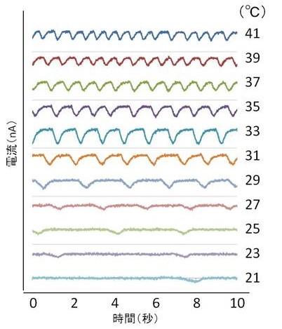
Figure 1 Changes in pulsation due to temperature changes
The number of beats and the time required for beats can be analyzed from the waveform of the measured current data.
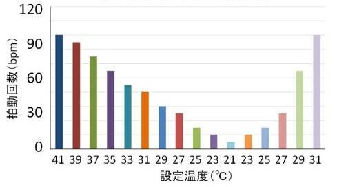
Figure 2 Beating number at each temperature
The beating frequency decreased by lowering the medium temperature and recovered with increasing temperature.
This data shows that temperature control during the assay is very important. An aluminum block warmer (HIENAI) is effective in preventing temperature drops during operations such as chemical addition and medium replacement .
*These data were provided by Mr. Hirano of the National Institute of Advanced Industrial Science and Technology.
References
- Yu Hirano, Mikie Kodama, Masahiro Shibuya, Yoshiyuki Maki and Yasuo Komatsu.
Analysis of beat fluctuations and oxygen consumption in cardiomyocytes by scanning electrochemical microscopy.
Anal Biochem, 2014, 447, 39-42. PMID: 24252541 DOI: 10.1016/j.ab .2013.11.008 - Sho Okumura, Yu Hirano, Yoshiyuki Maki and Yasuo Komatsu.
Analysis of time-course drug response in rat cardiomyocytes cultured on a pattern of islands.
Analyst, 2018,143, 4083-4089. PMID: 30083681 DOI: 10.1039/c8an01033a
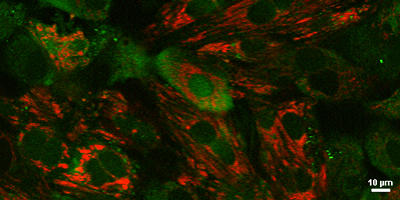
Imaging of calcium ions in rat myocardial cells
Photographing cooperation: Nikon Corporation
Imaging device: Confocal laser microscope system A1R (manufactured by Nikon Corporation)
Rat cardiomyocytes stained with two fluorescent dyes
Red: Mito-tracker Red (mitochondrial dye)
Green: Fluo-8 AM (calcium indicator)
Fluo-8 fluorescence changes in waves. Here is the video .
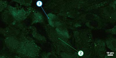
The fluorescence intensity of Fluo-8, a calcium indicator, was measured at two different points (① and ②) ( see video ).
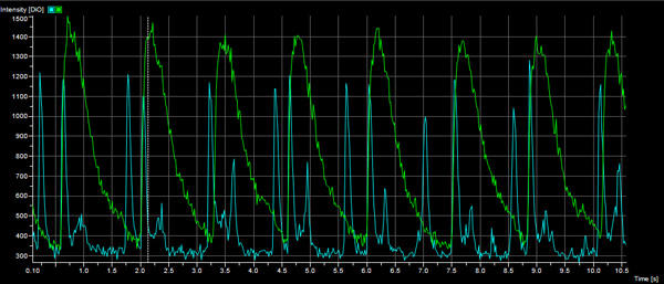
Above is a graph showing the fluorescence intensity at (1) for the blue line and (2) for the green line.
It became clear that the fluorescence intensity (calcium concentration) changed at a constant rhythm.
| Catalog Number | Product Name | Size |
| PMC-CMC12C | Cardiomyocytes (mouse) | 1 Vial |
| PMC-CMCM | Cardiomyocyte Culture Medium | 500 ml |