α-Synuclein aggregation assay kit
Intracellular recapitulation of α-Synuclein aggregate formation
The α-Synuclein Aggregation Assay Kit is a model that reproduces α-synuclein aggregate formation in cells, enabling in vitro screening of active ingredients.
*This product is a licensed product from Dr. Shigeto Hasegawa and Dr. Takashi Nonaka, Department of Dementia and Higher Brain Function Research, Tokyo Metropolitan Institute of Medical Science.
[Related information]
- Review article: Seed-dependent intracellular accumulation model of α-synuclein
- Dr. Takashi Nonaka, Dementia Project, Tokyo Metropolitan Institute of Medical Science - Alpha-synuclein antibodies [CAC]
- primarily for Parkinson's disease (PD), dementia with Lewy bodies (DLB), multiple system atrophy (MSA) research
Background
Dementia is a disease that impairs memory and judgment, and in Japan, where the elderly population is rapidly increasing, about 8 million people, including people at risk of dementia, are suffering. However, no radical cure has been found yet. Many years of research have shown that in many neurodegenerative diseases, including dementia, clusters (aggregates) of certain proteins are observed in degenerative sites such as the brain, and that the appearance of aggregates is closely related to the onset and progression of the disease. I have found that they are related.
Aggregates are accumulations of normal proteins that form abnormal structures in cells. In addition, the protein that accumulates in each disease is different. In the brains of patients with Parkinson's disease, dementia with Lewy bodies, and multiple system atrophy, α-synuclein is known to undergo structural changes in cells and form aggregates.
Composition
- This product provides enough reagent for 300 wells in a 24-well plate when prepared according to the preparation method described in the data sheet.
- The plasmid vector of this product uses a product synthesized by ATUM.
-
Content Amount Quantity pCMV-SNCA 32 μL 1 (α-synuclein expression plasmid vector, red cap) (concentration: 1.25 μg/μL) pCMV-NC 5 μL 1 (negative control vector, green cap) (Concentration: 1.25 μg/μL) pCMV-dGFP 5 μL 1 (dGFP expression plasmid vector, blue cap) (Concentration: 1.25 μg/μL) 20mM Tris-HCl Buffer 10 mL 1 (pH7.4) F-αSyn 32 μL 1 (α-synuclein fibrillating protein, yellow cap) (Concentration: 1 μg/μL) MultiFectam 0.33mg 1 (gene transfer reagent)
Things to prepare (Other necessary items)
- Assay cell line (recommended: SH-SY5Y)
- Culture medium (recommended: DE/F-12, 10% FBS, 1% NEAA)
- Opti-MEM® or serum-free medium (ThermoFisher Scientific: 31985062, etc.)
- Sterilized purified water (DNase, RNase free)
Experimental example for detection of synuclein aggregates
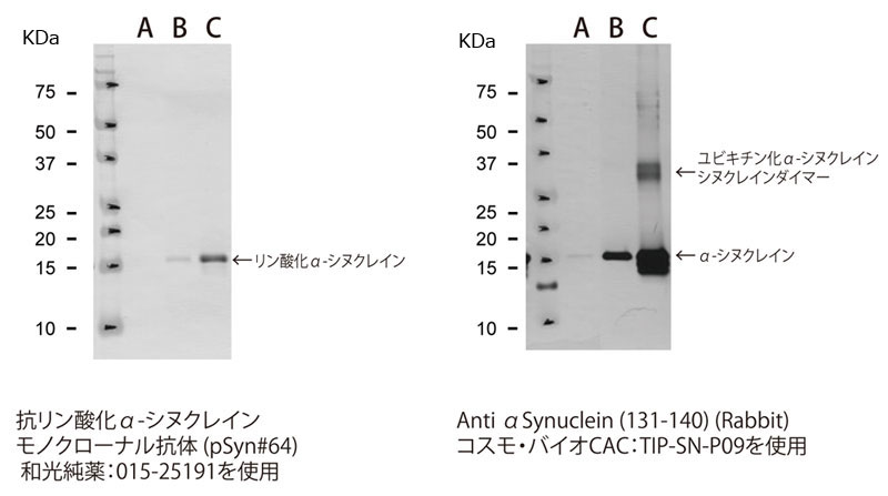
1. Evaluation by Western blotting
Western blot detection is possible using various synuclein antibodies.
Experimental conditions (example)
- Media removal
- Add 1 mL PBS to wells and collect cells by pipetting.
- Centrifuge at 4,500 rpm for 5 minutes and collect the cell pellet.
- 0.1 mL Lysis Buffer (10 mM Tris-HCl, pH 7.5 containing 0.8 M NaCl, 1 mM ethyleneglycol bis(2-aminoethyl ether)-N,N,N,N-tetraacetic acid (EGTA), 1 mM DTT and 1% N -Lauroylsarcosine sodium salt) and sonication
- Centrifuge at 50,000 rpm (100,000 xg) x 20 minutes, room temperature
- Add 20 μL 2 x SDS Buffer (0.125 M Tris-HCl pH6.8, 200 mM 1,4 dithiothreitol (DTT), 4% sodium dodecyl sulfate (SDS), 10% Sucrose, 0.01% BPB) to the pellet and sonicate. , 100 degrees, heat for 5 minutes, apply 5 μL
(click image to enlarge)
Fig. 1 Example of detection experiment of synuclein aggregates by Western blotting
A. pCMV-NC (negative control vector)
B. pCMV-SNCA (α-synuclein expression plasmid vector)
C. pCMV-SNCA + F-αSyn (α-synuclein introduction )
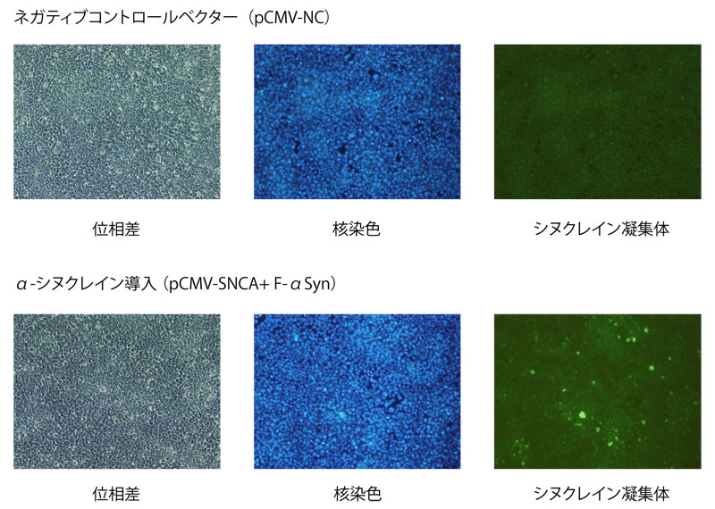
2. Detection using the Amyloid Structural Fluorescence Staining Kit
Double staining of aggregated α-synuclein and nuclei is possible using the Amyloid Structural Fluorescence Staining Kit (Cat. No. SYN02).
(click image to enlarge)
α-synuclein aggregation assay kit
| Catalog Number | Product Name | Size |
| CSR-SYN01 | Alpha-Synuclein Aggregation Assay Kit | 1 Kit |
Amyloid structure fluorescence staining kit
| Catalog Number | Product Name | Size |
| CSR-SYN02 | Amyloid Fluorescent Staining Kit | 1 Kit |
α-Synuclein fibrillating protein human/mouse
| Catalog Number | Product Name | Size |
| CSR-SYN03 | Alpha-Synuclein Fibrils, Human | 0.1 mg |
| CSR-SYN05 | Alpha-Synuclein Fibrils, Mouse | 0.1 mg |
Alpha-Synuclein Recombinant Protein Human/Mouse
| Catalog Number | Product Name | Size |
| CSR-SYN04 | Alpha-Synuclein, Recombinant, Human | 0.1 mg |
| CSR-SYN04 | Alpha-Synuclein, Recombinant, Human | 1 mg |
| CSR-SYN06 | Alpha-Synuclein, Recombinant, Mouse | 0.1 mg |
| CSR-SYN06 | Alpha-Synuclein, Recombinant, Mouse | 1 mg |