PD-L1/CD9 Exosome ELISA Kit (Human)
Cosmo Bio
- Catalog No.:
- HAK-HELPDL109-1
")
- Shipping:
- Calculated at Checkout
Product Description
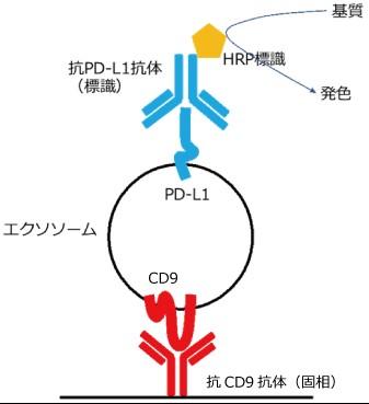
This product is a sandwich ELISA kit that uses high-performance antibodies against CD9, an exosome marker, and PD-L1, an immune checkpoint-related molecule, to detect PD-L1 molecules expressed on the surface of exosomes secreted by cells in human blood and cell culture fluid.
Background
What is PD-L1?
Cancer cells express PD-L1 (programmed cell death ligand-1) on their surface, which is a receptor for the PD-1 receptor expressed on the surface of activated T cells, and suppress T cell activity through PD-1/PD-L1 binding, thereby evading immune surveillance.1 ),2) In recent years, these immune surveillance mechanisms have been applied to cancer treatment, and anti-PD-1 and anti-PD-L1 antibodies have been developed to enhance T cell activation by inhibiting this binding, and their clinical efficacy and safety have been confirmed in various solid cancers.3 ),4) The
most well-known predictor of the efficacy of these immune checkpoint inhibitors is the expression of PD-L1 on cancer cells, and the PD-L1 IHC test (immunohistochemical staining method), which has been incorporated into clinical trials, is currently being developed to measure the PD-L1 expression rate in cancer tissues and cells, and is being used as a companion diagnostic.
In general, IHC testing is highly invasive and places a large burden on subjects, so research is also underway into liquid biopsies using exosomes that can reduce this burden.
PD-L1 is also expressed on exosomes, and has the same topology as PD-L1 on the surface of cancer cells, with the extracellular domain exposed on the surface of exosomes, which binds to PD-1 on T cells in a concentration-dependent manner5) . In addition, a comparison of circulating exosomes in the blood of metastatic melanoma patients and healthy subjects suggests that PD-L1 levels are significantly higher in metastatic melanoma patients5) . However, this method requires the purification of exosomes by ultracentrifugation, and direct measurement cannot be performed quickly and easily.
Features
- Highly sensitive detection of human PD-L1-positive exosomes by ELISA (detection sensitivity: 0.05 ng/mL * )
- Direct quantification of PD-L1-positive exosomes in human blood samples, cell culture supernatants, etc.
- No special equipment is required; measurements can be performed with a standard plate reader.
- Stability and reproducibility are ensured by using PD-L1/CD9 fusion protein (standard protein) instead of exosomes themselves, which lack storage stability as a standard reagent.
- Relative quantification of each sample is possible by reading it against a standard curve using PD-L1/CD9 fusion protein (standard protein)
*Calculated from the standard deviation of blank absorbance and the slope of the calibration curve6 )
Kit Components
| Contents | capacity | quantity |
|---|---|---|
| Anti-CD9 antibody immobilized plate | 96 well (8-well x 12 strips) | 1 sheet |
| Standard protein (PD-L1/CD9 fusion protein) | 100 µL | 1pc * |
| Assay Buffer | 25 mL | 1 bottle |
| Washing buffer (10x) | 25 mL | 1 bottle |
| HRP-labeled anti-PD-L1 antibody (500x) | 20 µL | 1 bottle |
| Substrate solution | 12 mL | 1 bottle |
| Stop solution ( 2N H2SO4 ) | 6 mL | 1 bottle |
| Plate seal | - | 2 sheets |
*; n=2, two calibration curves
Specification
| Cross-reactivity | Sample Type | Measurement range | sensitivity |
|---|---|---|---|
| Human | Serum, Plasma, Cell culture supernatant | 0.156–10 ng/mL | 0.05ng/mL |
Example data
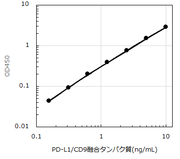
Figure 1. Standard curve
| Product Specifications | |
| Reactivity | Human |
| Documents & Links for PD-L1/CD9 Exosome ELISA Kit (Human) | |
| Datasheet | PD-L1/CD9 Exosome ELISA Kit Datasheet |
| Documents & Links for PD-L1/CD9 Exosome ELISA Kit (Human) | |
| Datasheet | PD-L1/CD9 Exosome ELISA Kit Datasheet |