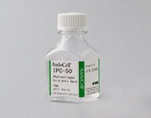
Atelocollagen, Bovine dermis, 5mg/mL

Cosmo Bio

SKU:
KOU-IPC-50
  • Atelocollagen, Bovine dermis, 5mg/mL
  • Three-dimensional culture of blood vessel endothelium using the collagen gel (Eguchi R, Hyogo College of Medicine Department of Environmental and Preventive Medicine)

An immunosuppressant FK506 is known to be involved in endothelial dysfunction inducing thrombotic microangiopathy after hematopoietic stem cell transplantation. In order to clarify the mechanism of the event, human umbilical vein endothelial cells (HUVEC) in collagen gel 3D culture were treated with FK506. In 3D culture, FK506 induced cell death and tube structure breakdown in a time- and concentration-dependent manner, but showed little effect on cells in monolayer culture. These results suggest 3D culture in collagen gel is useful to investigate in vivo phenomenon in vitro (Ref.6).
  • Comparison of albumin production of rat primary hepatocyte by various culture methods

1 uncoated
2 atelocollagen gel
3 native collagen gel
4 atelocollagen coated plate 5 native collagen coated plate 6 Company A’s coated plate 7 Company B’s coated plate 8 gelatin coated plate

Higher albumin production was maintained when cells were cultured on Atelocollagen and Native collagen gels ( 2 , 3 ) compared to other culture methods tested. In addition, results with both pre-coated plates ( 6 , 7 ) were similar to gelatin coated plates ( 8 ), suggesting that collagen degrades during storage.
  • Cell polarity formation of renal epithelial cells using collagen gel (Yonemura S, RIKEN Center for Life Science Technologies Ultrastructural Research Team)

Localization of ZO-1 (tight junction) and PKC Zeta (apical membrane) markers were observed inside spheroids of Madin-Darby canine kidney (MDCK) II cells cultured in collagen gel. On the other hand, localization of Scrib, a basal marker, was observed at the outer surface of spheroids. Thus, collagen gel 3D culture is effective for the formation of apical-basal polarity. Furthermore, collagen gel cultures showed a higher rate of single lumen formation compared to Matrigel and hanging drop culture (Ref. 4).
  • Invasion assay of cervical cancer cells in collagen gel (Uekita T, et al. National Defense Academy Division of Genome Biology)

Human cervical cancer cells (HCK1T-E cells) and active Ras transfected HCK1T-E cells (HCK1T-E-Hras cells) were seeded on collagen gel containing human fibroblast cells (Raft) to evaluate association between CDCP1 expression and cancer cell invasion. As a result, invasion of HCK1t-E-Hras cells into Raft was observed and remarkable expression of CDCP1 was detected in the invasion site. In addition, invasion into Raft was inhibited by CDCP1 siRNA transfection. Therefore, it was shown that the collagen gel was useful as a cancer cell invasion model to the submucosa (Ref. 3).
  • How to use Collagen Solutions
  • Molecular Structure of Atelocollagen and Native Collagen
  • Comparison list of the collagen solution products
  • Atelocollagen, Bovine dermis, 5mg/mL
Shipping:
Calculated at Checkout
$200.00
US Customers: This item requires a permit. Click here for more information.
Adding to cart… The item has been added

These products are prepared with highly purified type I collagen derived from bovine dermis. Neutral Solution products are pH neutral, come premixed in several commonly used culture media, and gelate at 37°C. In contrast to cell-derived soluble basement membrane preparations, Collagen Solutions are free from bioactive substances, nucreic acids, MMP, etc. allowing experimental results to be evaluated more clearly.

Applications

  • Collagen coating for cell cultureware
  • Cell culture in a collagen gel
  • Cell culture on a collagen gel

Features

  • Native collagen gelates when neutralized and placed under physiological condition.
  • Ready-to-use: atelocollagen is premixed with medium

This product is manufactured in Japan and produced from raw materials obtained from livestock certified born and raised in Australia or New Zealand. Japan, Australia and New Zealand are all recognized by the World Health Organization as having negligible risk for Bovine Spongiform Encephalitis (BSE). Nevertheless, please consult with your country's Customs office to confirm importability.

References

1. Arai KY, et al. Stimulatory eff ect of fi broblast-derived prostaglandin E2 on keratinocyte stratifi cation in the skin equivalent. (2014) Wound Repair
Regin. 22(6): 701-711
2. Sakakibara A, et al. Dynamics of centrosome translocation and microtubule organization in neocortical neurons during distinct modes of
polaristion. (2014) Cereb Cortex. 24(5): 1301-1310.
3. Uekita T, et al. Oncogenic Res/ERK signaling activates CDCP1 to promote tumor invasion and metastasis. (2014) Mol Cancer Res.12(10): 1449-
1459.
4. Yonemura S. Diff erential sensitivity of epithelial cells to extracellular matrix in polarity setabilishment. (2014) PLoS One. 9(11): e112922.
5. Correia AL, et al. The hemopexin domain of MMP3 is responsible for mammary epithelial invasion and morphogenesis through extracellular
interaction with HSP90 β . (2013) Genes Dev. 27(7): 805-817.
6. Eguchi R, et al. FK506 induces endothelial dysfunction through attenuation of Akt and ERK1/2 independently of calcineurin inhibition and the
caspase pathway. (2013) Cell Signal. 25(9): 1731-1738.


Documents & Links for Atelocollagen, Bovine dermis, 5mg/mL
Datasheet Atelocollagen, Bovine dermis, 5mg/mL Datasheet
Flyer Atelocollagen, Bovine dermis, 5mg/mL Flyer
Vendor Page Atelocollagen, Bovine dermis, 5mg/mL